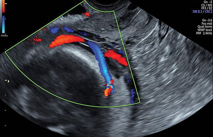
Ultrasound features suggestive of placenta accreta include deficiency of retroplacental sonolucent zone vascular lacunae myometrial thinning and interruption of the bladder line. The doctor recommended complete bed rest during the remainder of the womans pregnancy because of the abnormally.

Placenta Previa Obgyn Key

Placenta Previa Practical Approach To Sonographic Evaluation And Management

Placenta Previa Obstetrical Complications Due To Pregnancy Williams Manual Of Pregnancy Complications 23 Ed
One of the suggested mechanisms for threatened abortion is placental dysfunction which can also cause several late complications such as preeclampsia preterm labor preterm birth placental abruption placenta previa intrauterine growth.

Placenta previa sonography. Grayscale sonography has a sensitivity of 77 to 87 and a specificity of 96 to 98 for placenta accreta. Surgical puncture to aspirate fluid from the pelvic cavity. They are usually of no clinica.
They frequently occupy the potential space of the tunica vaginalis or sinus of the epididymis. Diagnostic ultrasound also called sonography or diagnostic medical sonography is an imaging method that uses high-frequency sound waves to produce images of structures within your body. The images can provide valuable information for diagnosing and treating a variety of diseases and conditions.
Clinical and Ultrasound Predictors of Placenta Accreta in Pregnant Women with Antepartum Diagnosis of Placenta Previa Women with a prior delivery by caesarean section have a high incidence of placenta accreta among women with antepartum diagnosis of placenta previa. Low-lying placenta occurs when the placenta extends into the lower uterine segment and its edge lies too close to the internal os of the cervix without covering itThe term is usually applied when the placental edge is within 05-50 cm of the internal cervical os 1Some alternatively give the term when the placental edge is within 2 cm from the internal cervical os 5. Scrotoliths also known as scrotal pearls are benign incidental extratesticular macrocalcifications within the scrotum.
The term pelvic sonography is defined as A. Because placenta previa may resolve close to term it is recommended that no decision on mode of delivery be made until after ultrasonography at 36 weeks25 Women whose placental edge is 2 cm or.

Pin On Obstetric Ultrasound
1

Comparison Of Transabdominal And Transvaginal Sonography In The Diagnosis Of Placenta Previa Petpichetchian 2018 Journal Of Clinical Ultrasound Wiley Online Library

Placenta Previa Transabdominal Sonography Shows The Cervix Lower Download Scientific Diagram
1

Placenta Previa Symptoms Ultrasound And Treatment
Classification Of Placenta Previa

Pdf Complete Placenta Previa Ultrasound Biometry And Surgical Outcomes Semantic Scholar
