Neck swellings are commonly encountered and present at all ages. Many of the disease states that affect the deep structures of the head and neck are confined.
Parapharyngeal Space Anatomy

Imaging Of The Parapharyngeal Space Sciencedirect

Other Neck Spaces Parapharyngeal Space Ranzcrpart1 Wiki Fandom
Imaging revealed that the mass in his right parapharyngeal carotid space had increased in size causing carotid stenosis.

Parapharyngeal space anatomy. The patient was hospitalized for 4 days and was treated with steroids. Your eustachian tube is located in the area known as parapharyngeal space. It runs from the front wall of the middle ear to the side wall of the nasopharynx.
Pericoronitis in two patients. Ranulas can be caused by trauma to the delicate sublingual gland ducts causing them. The infratemporal fossa is an irregularly shaped cavity that is a part of the skullIt is situated below and medial to the zygomatic archIt is not fully enclosed by bone in all directions.
In children the eustachian tube only slopes about 10 degrees downward. Pharyngeal superficial mucosal space. Radiological anatomy is crucial for radiologists and forms the base for learning radiology.
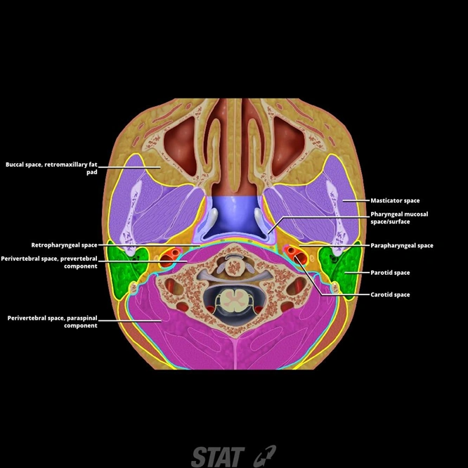
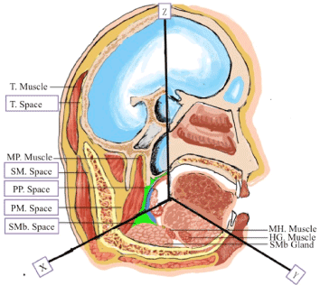
The deep anatomy is separated by fascial planes into seven deep compartments of the head and neck. IMAIOS and selected third parties use cookies or similar technologies in particular for audience measurement. It consists largely of fat neurovascular structures and in some definitions the retromandibular part of the deep lobe of the parotid gland.
Teeth are composed of multiple components. Anatomy and Development. A ranula is a type of mucocele mucous cyst that occurs in the floor of the mouth inferior to the tongue.
In the community inflammatory lymph nodes are most common while in the hospital environment the thyroid swelling or goiter is most frequently seen. Definitive treatment usually involves extraction of the involved tooth. Parapharyngeal Space Pharyngeal Mucosal Space Masticator Space Parotid Space Carotid Space Nasopharyngeal Perivertebral Space Paraspinal Perivertebral Space Prevertebral Buccal Space Parotid Nodes Labels OnOff.
It contains superficial muscles including the lower part of the temporalis muscle the lateral pterygoid muscle and the medial pterygoid muscleIt also contains important blood vessels. In their first year residents should be well versed with normal radiographs ultrasound and CT anatomy followed by MRI in the consequent years. Cookies allow us to analyze and store information such as the characteristics of your device as well as certain personal data eg IP addresses navigation usage or geolocation data unique identifiers.
A wide range of pathologic processes may involve the floor of the mouth the part of the oral cavity that is located beneath the tongue. It is the most common disorder associated with the sublingual glands due to their higher mucin content in secretions compared to other salivary glands. The differential diagnosis of a neck mass is extensive.
In adults the eustachian tube slopes downward about 35 degrees. Parapharyngeal or least commonly sublingual space. The parapharyngeal space also known as the prestyloid parapharyngeal space is a deep compartment of the head and neck around which most other suprahyoid fascial spaces are arranged.
Pharyngeal space infection most often arises via contiguous spread of infection from a peritonsillar or retropharyngeal abscess. They include lesions that arise uniquely in this location eg ranula submandibular duct obstruction as well as various malignancies inflammatory processes and vascular abnormalities that may also occur elsewhere in the head. The space between the roots is called the furcation F.
Other deep neck infections include retropharyngeal abscess and parapharyngeal space abscess also known as pharyngomaxillary or lateral pharyngeal space abscess.

Transoral Approach For Draining Parapharyngeal Space Abscesses Involving Multiple Maxillofacial Spaces
Esnr Org

Transcervical Approach For Removal Of Benign Parapharyngeal Space Tumors Sciencedirect

Percutaneous Biopsy Of Head And Neck Lesions With Ct Guidance Various Approaches And Relevant Anatomic And Technical Considerations Radiographics
:background_color(FFFFFF):format(jpeg)/images/article/en/the-para-and-retropharyngeal-spaces/2tEmtzKqjNappHRcfy7Vg_Pharynx.png)
Parapharyngeal And Retropharyngeal Spaces Anatomy Kenhub
Parapharyngeal Space Overview Radiology Key

References In Management Of Parapharyngeal And Retropharyngeal Space Infections Operative Techniques In Otolaryngology Head And Neck Surgery

The Parapharyngeal Space Review Of The Anatomy And Pathologic Conditions Semantic Scholar

